4. Determine the sectioning angles
Next, adjust the angles of the atlas slice to match the angles of sectioning. Even the best sectioning routines can induce small deviations from the vertical and horizontal planes. Furthermore, those angles can vary in a whole series, especially if the tissue was cut into two separate blocks. The cutting angles of the atlas should be adjusted to match the mediolateral and dorsoventral angles of the sections. This is done in the horizontal and sagittal navigation windows, respectively. Use either the rotation buttons (1) or rotation handles (2) to tilt the MRI template in the direction needed. Adjust the anteroposterior position to compensate for the rotation.
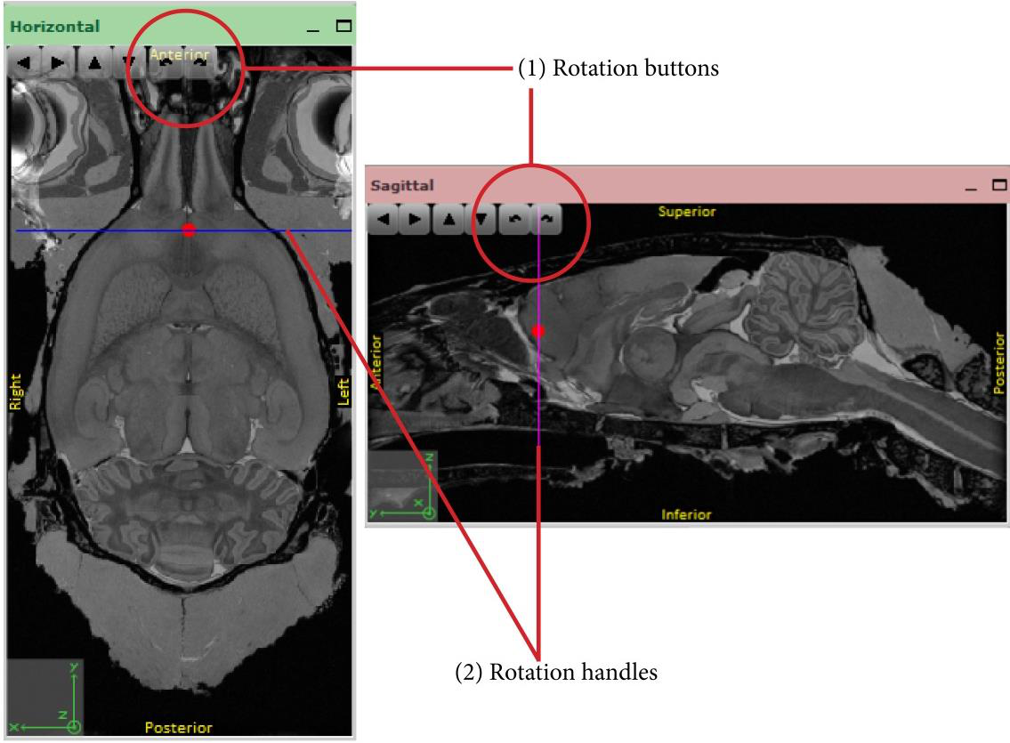
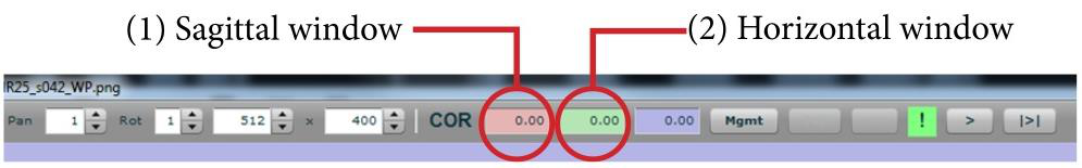
The angles of the current atlas slice relative to the default atlas plane can be read out in the boxes shown above, corresponding to the sagittal (1) and horizontal (2) navigation windows.
In coronal sections, the dorsoventral angle can be determined by examining the relationship between landmarks in dorsal and ventral parts of a section, e.g. between the corpus callosum and anterior commissure, between the dorsal and ventral hippocampus, or between the pons and inferior colliculus. The mediolateral angle can be determined by comparing landmark structures across hemispheres. It is most easily found by examining the development of the corpus callosum, anterior striatum, anterior commissure, anterior hippocampus, or size differences of the cortex in the posterior part of the brain.
Note: the results might look similar with angles that deviate 180 degrees (corresponding to looking at the animal from the back or from the front).